The Spinal Cord Is More Than a Pathway: A Deep Dive Into Its Structure and Function
The spinal cord is not just a conduction pathway but a segmental computational center that independently processes information and generates reflexes.
This article systematically outlines the spinal cord's organizational structure from an anatomical perspective. It is divided into 31 segments, with cervical and lumbar enlargements corresponding to limb control demands. In adults, the spinal cord ends at the L1 or L2 level; the lumbar cistern contains only cauda equina nerve roots suspended in CSF, explaining the low risk of lumbar puncture. Three meningeal layers, CSF, the filum terminale, and denticulate ligaments provide protection and stability. Spinal nerves formed by dorsal (sensory) and ventral (motor) roots create predictable dermatome maps for clinical localization. The dorsal horn processes sensory information, the ventral horn contains motor neurons as the final pathway for muscle contraction; the three white matter funiculi have distinct ascending and descending tracts. The core thesis: the spinal cord is both a passive conduction pathway and an active information-processing node, fundamental to understanding hierarchical control of behavior.
The spinal cord is often simplistically understood as a "cable" between the brain and the body, but this view falls far short of its true complexity. It is both an information highway and a computational center capable of independently processing information and generating reflex behaviors. This article takes an anatomical perspective to systematically outline the spinal cord's segmental organization, protective structures, nerve root distribution, and the fine division of labor within its internal gray and white matter.
I. Segmental Organization and Morphological Features
The spinal cord is a cylindrical neural structure that originates from the brainstem and begins at the foramen magnum, extending downward. It is divided into 31 segments: 8 cervical, 12 thoracic, 5 lumbar, 5 sacral, and 1 coccygeal. Each segment gives rise to a pair of spinal nerves that innervate the left and right sides of the body, connecting muscles, skin, joints, and internal organs.
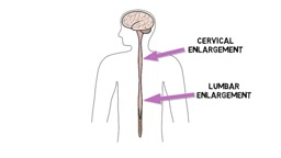
For most of its length, the spinal cord is only about one centimeter wide and gradually tapers downward. However, two regions are noticeably enlarged: the cervical enlargement and the lumbar enlargement. These enlargements correspond to an increased number of neurons controlling the upper and lower limbs—the more refined the limb control demands, the higher the local neuronal density.
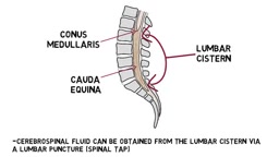
A common misconception is that the spinal cord runs through the entire vertebral canal. In adults, the spinal cord usually extends only to the level of the first or second lumbar vertebra, ending in a cone shape called the conus medullaris. This means that the vertebral canal below the lower lumbar region does not contain the spinal cord itself but rather consists of nerve roots issuing from lower segments.
II. Protective System: Meninges, Cerebrospinal Fluid, and Stabilizing Structures
Like the brain, the spinal cord is wrapped in three layers of connective tissue—the meninges. The outermost layer is the tough dura mater, beneath which lies the arachnoid mater, and the innermost layer is the pia mater, which closely adheres to the surface of the spinal cord. Between the arachnoid and pia mater is the subarachnoid space, filled with cerebrospinal fluid, which cushions mechanical impacts and maintains the physiological environment.
Below the conus medullaris, the subarachnoid space continues to form the lumbar cistern. The lumbar cistern contains no spinal cord, only the cauda equina nerve roots suspended in cerebrospinal fluid. This anatomical feature has direct clinical significance: lumbar puncture (commonly known as a "spinal tap") is usually performed here because it does not directly contact the spinal cord, significantly reducing the risk of injury.
The stability of the spinal cord also relies on two extensions of the pia mater: the filum terminale extends downward from the conus medullaris and attaches to the bottom of the vertebral canal, serving an anchoring role; the denticulate ligaments extend from both sides of the spinal cord and connect to the dura mater, working with the filum terminale to suspend and fix the spinal cord within the meninges.
Cerebrospinal fluid (CSF) is a colorless, transparent fluid produced mainly by the choroid plexus within the ventricles, circulating in the subarachnoid space around the brain and spinal cord. The total volume of CSF in adults is about 150 milliliters, with about half contained in the spinal subarachnoid space. CSF not only provides mechanical cushioning but also performs functions such as nutrient transport, metabolic waste clearance, and regulation of intracranial pressure. Because CSF has a lower density than spinal cord tissue, the spinal cord parenchyma is suspended in the subarachnoid space, further reducing mechanical stress during daily activities.
Lumbar puncture is anatomically based on the fact that the lumbar cistern is the widest region of the subarachnoid space without spinal cord parenchyma. The puncture needle typically enters at the L3-L4 or L4-L5 interspace, passes through the dura mater and arachnoid mater to reach the lumbar cistern, and collects CSF for diagnosis (e.g., cell count, protein quantification, pathogen detection). Because the cauda equina nerve roots float in the CSF, they can be pushed aside by the needle, so the risk of injury is extremely low.
III. Nerve Roots and Dermatomes: Segmental Functional Mapping
Each spinal cord segment gives rise to a pair of nerve roots on each side: the dorsal root and the ventral root. The dorsal root carries sensory information into the spinal cord; at its entry point is an enlarged structure called the dorsal root ganglion, which contains the cell bodies of primary sensory neurons. The ventral root carries motor commands out of the spinal cord to target organs such as muscles.
The dorsal and ventral roots merge to form the spinal nerve. Spinal nerves are mixed nerves, containing both sensory and motor fibers. This segmental organization forms a predictable body surface map—the dermatome, which is the area of skin primarily innervated by the sensory fibers of a given spinal nerve. There is some overlap between adjacent dermatomes, but dermatome maps remain clinically valuable: by observing the distribution pattern of sensory loss, numbness, tingling, or pain, doctors can infer the damaged spinal nerve or spinal cord segment.
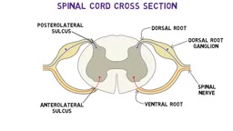
The dorsal root ganglion (DRG) is a cluster of sensory neuron cell bodies located at the dorsal root entry zone. These neurons are pseudounipolar: their peripheral processes extend to the skin, joints, and internal organs, while their central processes enter the dorsal horn of the spinal cord. The DRG is the first station for sensory signals entering the central nervous system and is also a site where several viruses (such as the varicella-zoster virus) can lie dormant.
The clinical value of the dermatome map lies not only in localizing the damaged segment but also in distinguishing radicular from peripheral nerve lesions. For example, the L5 dermatome covers the lateral lower leg and dorsum of the foot; if sensation is lost in this area while adjacent dermatomes are normal, it suggests L5 nerve root compression (e.g., disc herniation). There is a 1-2 segment overlap between adjacent dermatomes, so a single nerve root injury usually does not cause complete sensory loss but rather presents as hypoesthesia or paresthesia.
IV. Gray Matter and White Matter: Internal Functional Zoning
The most prominent feature of the spinal cord cross-section is the distribution of gray and white matter. Gray matter is located centrally, shaped like a butterfly or the letter H, and is surrounded by white matter. Gray matter is rich in neuronal cell bodies, dendrites, synapses, and unmyelinated axons; white matter consists mainly of myelinated axons, whose white color comes from the lipid insulating layer of myelin.
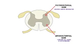
Gray matter is divided into three horn regions. The dorsal horn (posterior horn) receives a large amount of incoming sensory information, processing touch, pressure, pain, temperature, and proprioception. The ventral horn (anterior horn) contains motor neuron cell bodies whose axons leave the spinal cord to innervate skeletal muscles; it is the final pathway for the nervous system to produce muscle contraction. The intermediate gray matter, located between the dorsal and ventral horns, participates in sensory integration, motor coordination, and autonomic regulation.
White matter is divided into three funiculi: the dorsal funiculus, lateral funiculus, and ventral funiculus. The dorsal funiculus contains the dorsal columns, which carry fine touch, vibration, pressure, and conscious proprioception upward. The lateral funiculus contains both ascending and descending tracts: the spinothalamic tract transmits pain and temperature information; the spinocerebellar tract sends proprioceptive information to the cerebellum to assist motor coordination and posture maintenance; the lateral corticospinal tract is the main pathway for voluntary movement, originating in the cerebral cortex and controlling fine skilled movements of the limbs. The ventral funiculus contains the anterior corticospinal tract, which participates in trunk and upper limb muscle control, as well as pathways related to posture, balance, and reflex movements, such as the vestibulospinal and reticulospinal tracts.
The lateral corticospinal tract is the core pathway for voluntary motor control. It originates from pyramidal cells in the primary motor cortex (Brodmann area 4) and premotor areas, descends through the internal capsule, cerebral peduncle, and basilar pons, and at the medullary pyramidal decussation about 85% of fibers cross to the opposite side, forming the lateral corticospinal tract in the lateral funiculus of the spinal cord. This tract mainly controls fine skilled movements of distal limb muscles (such as independent finger movements). The uncrossed fibers form the anterior corticospinal tract, which descends in the ipsilateral ventral funiculus and mainly innervates trunk and proximal limb muscles.
The spinothalamic tract transmits pain, temperature, and crude touch. Its fibers come from contralateral dorsal horn neurons (second-order sensory neurons), cross in the anterior white commissure, and ascend to the thalamus. This tract is arranged in layers within the spinal cord: fibers from the sacral segments are located most laterally, and cervical fibers most medially. This arrangement helps clinically localize intramedullary spinal cord lesions (e.g., when syringomyelia expands from the center outward, it first affects cervical fibers and later sacral fibers).
V. Understanding Beyond the "Pathway"
The complexity of the spinal cord is reflected at two levels. Longitudinally, the 31 segments form precise correspondences with specific body regions; transversely, the gray matter horns and white matter tracts each have their own roles. More critically, local circuits within the spinal cord can process information and generate reflex behaviors without direct involvement of the brain. This means the spinal cord is both a passive conduction pathway and an active information-processing node. Understanding this dual attribute is fundamental to understanding how the nervous system controls behavior hierarchically.
One-line Summary
The spinal cord is not only a conduction pathway but also a segmental computational center capable of independently processing information and generating reflexes.
Paragraph Summary
This article systematically outlines the organizational structure of the spinal cord from an anatomical perspective. The spinal cord is divided into 31 segments, with the cervical and lumbar enlargements corresponding to limb control demands. In adults, the spinal cord ends at the level of the first or second lumbar vertebra; the lumbar cistern contains only cauda equina nerve roots suspended in cerebrospinal fluid, which is why lumbar puncture carries low risk. Three meningeal layers, cerebrospinal fluid, the filum terminale, and denticulate ligaments together provide protection and stability. Spinal nerves are formed by the merger of dorsal roots (sensory) and ventral roots (motor), creating predictable dermatome maps used for clinical localization of lesions. The dorsal horn of gray matter processes sensory information, while the ventral horn contains motor neurons and is the final pathway for muscle contraction; the three funiculi of white matter each have distinct ascending and descending tracts. The core thesis is: the spinal cord is both a passive conduction pathway and an active information-processing node, and understanding its dual attributes is fundamental to understanding how the nervous system controls behavior hierarchically.
Related articles
 Tech Frontiers
MuJoCo: An Analysis of DeepMind's Open-Source General-Purpose Physics Simulator
An in-depth analysis of Google DeepMind's open-source physics simulator MuJoCo: high-performance C++ kernel, MJCF modeling format, contact solving principles, MJX GPU acceleration, and robot reinforcement learning use cases.
 Tech Frontiers
Claude API Quick Start: The Official Quickstarts Project Collection
Anthropic's official claude-quickstarts project collection helps developers quickly build deployable apps with the Claude API. Nearly 20k stars, Python-based, great for beginners and prototyping.
 Tech Frontiers
AI Agents Connecting Directly to APIs: Which Calls Can Be Safely Allowed?
AI agents can call any API, but the real challenge is deciding which calls can be safely allowed. This article analyzes allowance decisions from three dimensions—intent matching, least privilege, and controllable impact—and argues that the control layer should sit outside the agent, combined with audit traceability to build a trustworthy API call governance mechanism.