OpenRadView: A Free, Open-Source Medical Imaging Viewer with CT/MRI Support

OpenRadView is a free, open-source tool for viewing CT, MRI, and X-ray images locally, not for clinical diagnosis.
OpenRadView is a free, open-source medical imaging viewer featured on Product Hunt that opens DICOM and NIfTI files from scan discs or archives directly on a personal computer. It offers linked axial, coronal, and sagittal views, an experimental 3D view, basic measurement tools, and an optional MCP interface for connecting your own AI agent. It is aimed at personal research, education, and preliminary understanding of imaging content, is not clinically validated, and explicitly requires users to consult a qualified doctor for interpretation.
OpenRadView is a newly launched free and open-source tool featured on Product Hunt that lets ordinary people open DICOM and NIfTI files from CT, MRI, and X-ray scans on their own computers. It is not meant to replace doctors, but rather to provide a low-barrier entry point for personal research, education, and a preliminary understanding of imaging content.
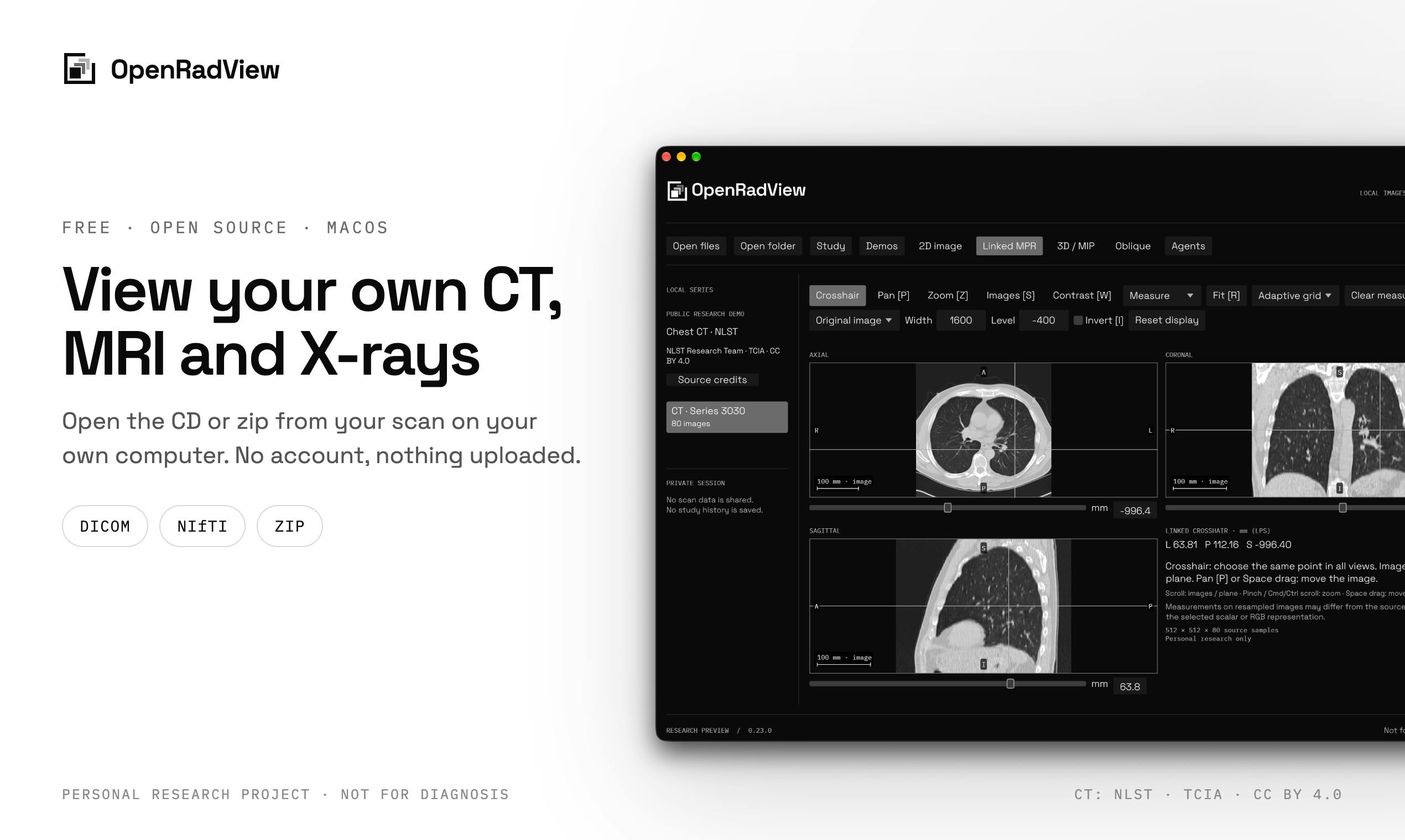
From Scan Discs to Local Viewing: What OpenRadView Can Do
After a patient undergoes a CT or MRI, they typically receive a disc or compressed archive containing the imaging data. These files are mostly in DICOM format, which ordinary software struggles to open directly. OpenRadView's core value is turning this data from something you can only read reports about into something you can browse yourself.
It supports reading DICOM and NIfTI files directly from scan discs or zip archives and rendering them on a local computer. The viewing features include linked axial, coronal, and sagittal views—these three perspectives are synchronized to the same position, making it easier for users to understand the spatial location of lesions or structures. There is also an experimental 3D view and measurement tools, allowing users to take basic length or angle measurements on images.
These features are common in professional radiology workstations, but OpenRadView makes them free, open-source, and personally accessible—which is the main reason it has attracted attention.
DICOM (Digital Imaging and Communications in Medicine) is the international standard used in the medical imaging field, specifying how imaging data is stored, encoded, and transmitted. A single CT or MRI examination often produces hundreds to thousands of slices, each carrying not only pixel data but also metadata such as patient information, equipment parameters, slice thickness, and position—all packaged in DICOM format. It is essentially a container of "images + tags," which is why ordinary image viewers cannot parse it directly.
NIfTI (Neuroimaging Informatics Technology Initiative) is a format commonly used in neuroimaging research, mostly for analyzing and processing MRI data. It typically stores three-dimensional volume data as a single file with accompanying spatial coordinate information. Its difference from DICOM is that it is geared more toward scientific analysis workflows rather than native output from hospital equipment. OpenRadView supports both formats, meaning it serves both the original disc data that patients receive and the analysis files used in research and teaching.
Axial, coronal, and sagittal are the three standard anatomical planes in medical imaging: axial is the "horizontal slice" cutting across the body, coronal is the "frontal slice" cutting from front to back, and sagittal is the "side slice" cutting from left to right. Any three-dimensional image can be browsed slice by slice along these three directions, and linked views synchronize all three planes to the same spatial point, helping users build a three-dimensional sense of orientation. This is also the basic way radiologists read scans, and OpenRadView replicates it on a personal computer.
Aimed at Personal and Educational Scenarios, With a Clear Clinical Boundary
OpenRadView's positioning is very clear: a personal research project, for educational purposes, not clinically validated. The product page explicitly states: "Ask a qualified clinician to interpret your scans; do not use the app for decisions about care."
This positioning is commendable. The interpretation of medical images relies heavily on professional training and clinical context, and ordinary users viewing images on their own can easily misread or over-interpret them. OpenRadView gives the tool to users while leaving interpretation to doctors—a sense of boundaries that is especially important in health-related tools.
It also offers an MCP (Model Context Protocol) access option, allowing users to connect their own AI agent. This means technically savvy users can explore the possibility of using AI to assist in understanding imaging data, but the output should likewise be treated with caution.
The Dual Appeal of Open Source and AI Integration
OpenRadView is released as open source, with code hosted on GitHub, and its category tags include Education, Artificial Intelligence, and Medical. This reflects a current trend: medical imaging tools are extending from closed professional systems toward open-source, programmable, and integrable directions.
MCP support is one of the more forward-looking design choices. MCP is a protocol that lets AI models interact with external tools and data sources, and OpenRadView allows users to connect their own AI agent to the image viewing workflow, potentially for automated annotation, structured descriptions, or assisted retrieval. However, the product page does not elaborate on the specific capability boundaries of MCP—this part remains experimental exploration.
It is worth noting that no AI interpretation of medical images can replace clinical diagnosis. OpenRadView's MCP feature is better suited for developers and researchers doing technical validation, rather than for patients judging their own conditions.
MCP (Model Context Protocol) is an open protocol proposed by Anthropic for letting large language models call external tools and read data sources in a standardized way. You can think of it as a "universal plug" between AI applications and the outside world: AI agents discover and call available tools through MCP servers without having to write separate integration code for each data source. OpenRadView's MCP access means that a user's AI agent can theoretically read the state of the image viewer or trigger operations for automated workflows. But the protocol itself only defines how interactions occur; it does not guarantee that the AI understands the image content accurately—which is why the product labels it as an experimental feature.
Who Is It For?
OpenRadView is best suited to three groups: ordinary users who are curious about their own images and want an intuitive understanding of what was scanned; students and teachers in medical education who can use it to demonstrate anatomical structures and imaging orientations; and developers and researchers who can use the open-source code and MCP interface for technical experiments related to medical imaging.
For patients who need a clinical diagnosis, the right approach is still to bring the imaging materials to a qualified doctor. OpenRadView is a viewing tool, not a diagnostic tool. Its value lies in lowering the barrier to accessing medical imaging data, not in replacing professional judgment.
One-line Summary
OpenRadView is a free, open-source tool that lets individuals view CT, MRI, and X-ray images locally, but it is explicitly not intended for clinical diagnosis.
Paragraph Summary
OpenRadView is a free, open-source medical imaging viewing tool featured on Product Hunt that supports opening DICOM and NIfTI files directly from scan discs or archives on a personal computer. It offers linked axial, coronal, and sagittal views, an experimental 3D view, and basic measurement tools, plus an optional MCP interface for connecting a user's own AI agent. Its positioning is clear: aimed at personal research, education, and preliminary understanding of imaging content, not clinically validated, and it explicitly asks users to have a qualified doctor interpret results and not to use it for medical decisions. Its value lies in lowering the barrier to accessing medical imaging data, making it suitable for curious ordinary users, medical education scenarios, and developers conducting technical experiments.
Related articles

The Truth About AI Coding: Code Output Soars, So Why Is Delivery Still Hard to Speed Up?
Engineering leaders from Databricks, Uber, and Google Maps discuss AI coding's real impact: code output soars but delivery bottlenecks shift, code review faces rethinking, and enterprises begin fine-grained token governance.

A New Paradigm for Text Editing in Compose: TextFieldState Replaces the Old API
Jetpack Compose introduces the TextFieldState API to fix dropped characters and cursor jumps from async input. The old value/onValueChange callbacks will be deprecated in 1.13. This article covers InputTransformation, OutputTransformation, four state hoisting patterns, and new formatted text editing capabilities.

Remote Compose: A New Document-Driven Paradigm for Cross-Platform UI on Android
Remote Compose transmits compact binary documents instead of screen pixels, enabling efficient UI rendering on remote surfaces like Wear OS and widgets. This article covers its document creation API, expression animations, size optimization techniques, and Wear OS progress.